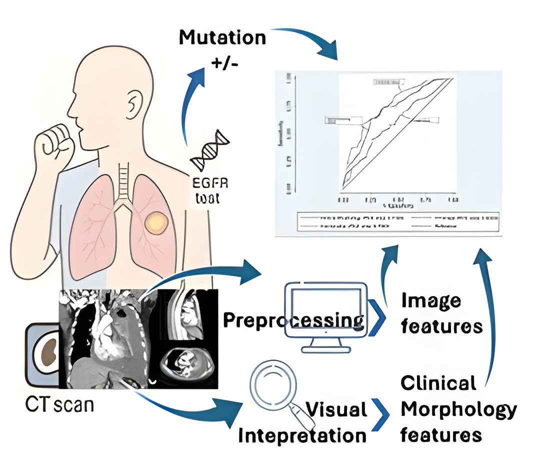
Tyrosine kinase inhibitors (TKIs) have demonstrated favorable responses in lung adenocarcinoma patients with EGFR (Epidermal Growth Factor Receptor) mutations, leading to improved 5-year progression-free survival rates. However, identifying EGFR mutation status typically requires molecular testing through biopsy, which is invasive and may yield insufficient tissue samples. For decades, computed tomography (CT) scans have served as the primary imaging modality in the evaluation and management of lung adenocarcinoma. Radiological interpretation of CT images provides valuable information regarding tumor characteristics, including morphology and invasiveness. Additionally, CT imaging offers high spatial resolution and distinct pixel-based features that can be further analyzed for quantitative assessment. Given these advantages, there is a growing need to develop non-invasive biomarkers to predict EGFR mutation status. This study aimed to develop a predictive model incorporating both clinical-morphological characteristics and CT image features. A retrospective cohort observational study was conducted using data from 179 patients diagnosed with lung adenocarcinoma who underwent CT scanning and EGFR molecular testing between 2018 and 2023 at Saiful Anwar General Hospital, East Java, Indonesia. Clinical variables included age and sex, while morphological features from CT reinterpretation included tumor size, ground-glass opacity, interstitial thickening, tumor margins, air bronchogram, necrosis, emphysema, nodules, lymph node involvement, metastasis, and pleural involvement. The combined model demonstrated superior performance compared to individual feature sets, achieving a sensitivity of 54.76%, specificity of 82.24%, and an AUC of 0.76 (95% CI: 0.69–0.83). These findings suggest that integrating clinical-morphological and CT imaging features improves the prediction of EGFR mutation status.