aimachine-learningmedical-imagingmedicinenlp
medical-imaging

Nature Communications

Biological sciences : Scientific Reports subject feeds
aideep-learningmachine-learningmedical-imagingmedicine
Biological sciences : Scientific Reports subject feeds
aimachine-learningmedical-imagingmedicine
Biological sciences : Scientific Reports subject feeds
aideep-learningmedical-imagingmedicine

The Medical News
diagnosticsmedical-imagingmedicine

ODU Digital Commons

educationmedical-imagingmedicine
Biological sciences : Scientific Reports subject feeds
aideep-learningmedical-imagingmedicine
Biological sciences : Scientific Reports subject feeds
aideep-learningmedical-imagingmedicine

Frontiers in Artificial Intelligence | New and Recent Articles
aimachine-learningmedical-imagingmedicine
Frontiers in Artificial Intelligence | New and Recent Articles
Aneta Lisowska
17d ago
aimachine-learningmedical-imagingmedicinenlp

3D Printing Industry

diagnosticsmedical-imagingmedicine

Frontiers in Neuroscience | New and Recent Articles
Ganqin Du
20d ago
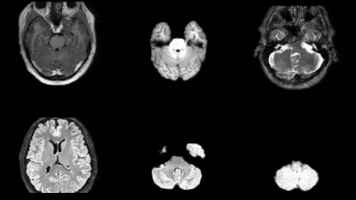
aialzheimer-s-diseasedeep-learningmedical-imagingmedicine

Frontiers in Reproductive Health | New and Recent Articles
Rajat Thomas
20d ago
aicomputer-visionmachine-learningmedical-imagingmedicine
Frontiers in Artificial Intelligence | New and Recent Articles
Costinela Corciu
20d ago
aideep-learningmedical-imagingmedicine

Agentic AI / Generative AI – NVIDIA Technical Blog
aideep-learningmachine-learningmedical-imagingmedicine
The Medical News
aimachine-learningmedical-imagingmedicine

Biological sciences : Scientific Reports subject feeds
Xiang Liu et al.
5/15/2026
aimachine-learningmedical-imagingmedicine

Frontiers in Computational Neuroscience | New and Recent Articles
Tariq Sadad
5/13/2026
diagnosticsmedical-imagingmedicine

Newswise: Latest News
Department of Energy·Office of Science
5/13/2026
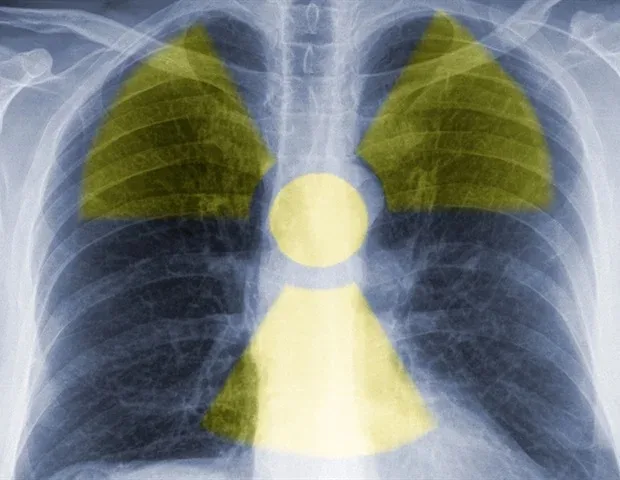
cancer-treatmentmedical-imagingmedicineoncologytechnology

Acoustics.org
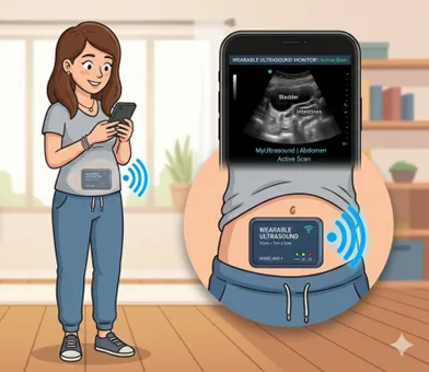
medical-imagingmedicinetechnologywearables
Sign up to keep scrolling
Create your feed subscriptions, save articles, keep scrolling.
Already have an account?